Online first
Bieżący numer
Archiwum
O czasopiśmie
Polityka etyki publikacyjnej
System antyplagiatowy
Instrukcje dla Autorów
Instrukcje dla Recenzentów
Rada Redakcyjna
Komitet Redakcyjny
Recenzenci
Wszyscy recenzenci
2024
2023
2022
2021
2020
2019
2018
2017
2016
Kontakt
Bazy indeksacyjne
Klauzula przetwarzania danych osobowych (RODO)
OPIS PRZYPADKU
Diagnozowanie ADHD z zastosowaniem QEEG i planowanie terapiiEEG-biofeedback (neurofeedback) – badania pilotażowe
1
Uniwersytet Rzeszowski, Polska
2
ADEA LTD, Bulgaria
Autor do korespondencji
Med Og Nauk Zdr. 2021;27(2):205-212
SŁOWA KLUCZOWE
DZIEDZINY
STRESZCZENIE
Wprowadzenie:
Wśród przyczyn zespołu nadpobudliwości psychoruchowej z deficytem uwagi (ADHD) wymienia się głównie uwarunkowania biologiczne (genetyczne, biochemiczne oraz anatomiczne). Czynniki środowiskowe wpływają również w istotny sposób na funkcjonowanie jednostki, ale są w dużej mierze konsekwencją zdeterminowanych biologicznie specyficznych reakcji dziecka czy osoby dorosłej z ADHD. Zatem wczesne diagnozowanie dysfunkcji stanowi istotny warunek podjęcia właściwych oddziaływań, sprzyjających złagodzeniu określonych objawów i uniknięciu różnych konsekwencji związanych z deficytem uwagi i nadruchliwością. W diagnozowaniu niezbędne jest uwzględnienie obiektywnych i subiektywnych symptomów zaburzeń. Przydatne są tu również: wywiad, obserwacja, a także testy psychologiczne. Ze względu na trudności w diagnozie różnicowej związanej z różnego typu schorzeniami istotne są również badania na-tury biologicznej. Szczególnie pomocne w diagnozowaniu jest ilościowe badanie EEG, czyli QEEG.
Cel pracy:
Analiza przydatności diagnozowania osób z ADHD metodą QEEG, która jest również podstawą do planowania wspomagającej terapii EEG-biofeedback.
Materiał i metody:
Badania mają charakter studiów przypadków. W pracy przedstawiono pogłębioną analizę wyników QEEG pięciorga losowo wybranych dzieci z ADHD
Wyniki:
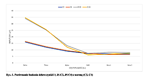
Badanie dzieci z ADHD przy pomocy QEEG wskazuje na powtarzające się zależności, występowanie wysokich am-plitud fal niskoczęstotliwościowych – delta, theta, alfa oraz niskie amplitudy fal wysokoczęstotliwościowych w porównaniu do niskoczęstotliwościowych. Stwierdzono natomiast zbliżone amplitudy fal wysokoczęstotliwościowych – SMR, beta 1 i beta 2 oraz niższe amplitudy fal beta 2 od alfa i theta.
Wnioski:
Badanie QEEG jest bardzo pomocne w diagnozowaniu ADHD. Metoda ta dostarcza istotnych informacji w plano-waniu terapii EEG-biofeedback i sprawdzaniu jej skuteczności.
Introduction:
Among the causes of attention deficit hyperactivity disorder (ADHD) are mentioned mainly biological conditions (genetic, biochemical and anatomical). Environmental factors also significantly affect the functioning of the individual, but are largely a consequence of the determined biologically specific reactions of a child or an adult with ADHD. Thus, early diagnosis of dysfunctions is an important precondition for undertaking appropriate actions to mitigate specific symptoms and avoid various consequences related to attention deficit and hyperactivity. In diagnosing, it is necessary to take into account the objective and subjective symptoms of disorders. Here, an interview, observation, and psychological tests are also useful. Due to the difficulties in differential diagnosis related to various types of diseases, biological research is also important. Quantitative EEG (QEEG) is particularly helpful in making a diagnosis.
Objective:
The aim of the study was analysis of the usefulness of the QEEG method in diagnosing persons with ADHD, which is also the basis for planning supportive EEG biofeedback therapy.
Material and Methods:
The research presents a case study, which includes an in-depth analysis of the QEEG results concerning five randomly selected children with ADHD.
Results:
The QEEG assessment of children with ADHD shows repeated dependencies, the presence of high amplitudes of low-frequency waves – Delta, Theta, Alpha, and low amplitudes of high-frequency waves, compared to low-frequency waves. However, similar amplitudes of high-frequency waves were found – SMR, Beta1 and Beta2, and lower amplitudes of Beta2 waves, compared to Alpha and Theta.
Conclusions:
The QEEG test is very helpful in diagnosing ADHD. This method provides important information while planning EEG biofeedback therapy and checking its effectiveness.
Kopańska M, Ochojska DB, Dejnowicz-Velitchkov A. Diagnozowanie ADHD z zastosowaniem QEEG i planowanie terapii EEG-biofeedback (neurofeedback) – badania pilotażowe. Med Og Nauk Zdr. 2021; 27(2): 205–212. doi: 10.26444/monz/131993
REFERENCJE (39)
1.
Thapar A, Cooper M, Eyre O, et al. Practitioner review: What have we learnt about the causes of ADHD. J Child Psychol. Psychiatry. 2013; 5 4 (1): 3 –13.
2.
Gaidanowicz R, Deksnyte A, Palinauskaite K, et al. ADHD – plaga XXI wieku? Psychiatr Pol. 2018; 52(2): 287–387.
3.
De La Fuente A, Xia S, Branch C, Li X. A review of attention-deficit/hyperactivity disorder from the perspective of brain networks. Front Hum Neurosci. 2013; 7: 192.
4.
Rhodes SS. Parenting dependent young adults with ADHD. Pediatr Nurs. 2017; 43(5): 243.
5.
Philipp-Wiegman F, Retz-Junginger P, Retz W, Rosler M. The intrain-dividual impact of ADHD on the transition of adulthood to old age. Eur Arch Psychiatry Clin Neurosci. 2016; 266: 367–369.
6.
Wasserstein J. Diagnostic issues for adolescent and adults with ADHD. J Clin Psychol. 2005; 61: 535–547.
7.
Ochojska D, Pasternak J. Postawy rodzicielskie w percepcji studentów z ADHD. Wychowanie w Rodzinie. 2019: 20(1): 180–194.
8.
Munden A, Arcelus J. ADHD. Nadpobudliwość ruchowa. Kraków: Bellona; 2006: 130–133.
9.
Hetmańczyk H, Kawiak E. Diagnozowanie zespołu nadpobudliwości psychoruchowej z wykorzystaniem testu MOXO. Edukacja – Technika – Informatyka. 2018; 23(1): 237–241.
10.
Pisula A, Bryńska A, Kołakowski A, et al. ADHD – zespół nadpobud-liwości psychoruchowej. Przewodnik dla rodziców i wychowawców. Gdańsk: GWP; 2018.
11.
Orylska A, Jagielska G. Diagnoza zespołu nadpobudliwości psychoru-chowej z deficytem uwagi u dzieci w wieku przedszkolnym. Psychiatr Psychol K lin. 2011; 11(2): 115 –119.
12.
Conners CK, Sitarenios G, Parker JD, et al. The Revised Conners’ Pa-rent Rating Scale (CPRS-R): factor structure, reliability, and criterion validity. J Abn Child Psychol. 1998; 26: 257–268.
13.
Conners CK. Conners Early Childhood Manual. Multi-Health Systems: New York; 2009.
14.
Bober-Płonka B, Kuleta-Krzyszkowiak M, Wasilewska M. Funkcjono-wanie studentów z ADHD – różnice między mężczyznami i kobietami. Edukacja – Technika – Informatyka. 2019; 1: 11–20.
15.
Kooij JJS, Francken MH. Diagnostic Interview for ADHD adults (Diva 2.0). In: Kooij JJS. Adult ADHD. Diagnostic Assessment and treatment. New York: Springer; 2013. p. 97–99.
16.
Levy E, Traicu A, Iyer S, et al. Psychotic disorders comorbid with attention-deficit hyperactivity disorder. An important knowledge gap. Can J Psychiatry. 2015; 60 (3 suppl. 2): 48–52.
17.
Blom JD, Niemantsverdriet M, Spuijbroek A, et al. Attention Deficit Disorder Psychosis. In: Sharpless BA, editor. Unusual and rare psy-chological disorders. A Handbook for clinical practice and research. Oxford: Oxford University Press; 2017: 78–81.
18.
Faraone CV, Biederman J, Spencer T, et al. Diagnosing adult attention deficit hyperactivity disorder: are late onset and subthreshold diagnosis valid? Am J Psychiatry. 2006; 163(10): 1720–1729.
19.
Lipowska M, Dykalska-Bieck D. Czy impulsywność w ADHD ma kom-ponenty temperamentalne? Psychiatr Psychol Klin. 2010; 10(3): 16–181.
20.
Fetz EE. Volitional control of neural activity: Implications for brain--computer interfaces. J Physiol. 2007; 15: 571–579.
21.
Walkowiak H. EEG biofeedback: charakterystyka, zastosowanie, opinie specjalistów. Studia Edukacyjne. 2015; 36: 307–325.
22.
Barry RJ, Clarke AR, Johnstone SJ, et al. EEG differences between eyes--closed and eyes-open resting conditions. Clin Neurophysiol. 2007; 118: 2765–73.
23.
Billeci L, Sicca F, Maharatna K, et al. On the application of quantitative EEG for characterizing autistic brain: a systematic review. Front Hum Neurosci. 2013; 7: 442.
24.
Chen C, Zhou C, Cavanaugh JM, et al. Quantitative electroencepha-lography in a swine model of blast-induced brain injury. Brain Inj. 2017; 31: 120–126.
25.
Wiśniewska M, Gmitrowicz A, Pawełczyk N. Zastosowanie QEEG w psychiatrii z uwzględnieniem populacji rozwojowej. Psychiatr Psychol Klin. 2016; 16: 188–193.
26.
Caviness JN, Hentz JG, Belden CM, et al. Longitudinal EEG changes correlate with cognitive measure deterioration in Parkinson’s disease. J Parkinsons Dis. 2015; 5: 117–24.
27.
Caviness JN, Utianski RL, Hentz JG. Differential spectral quantitative electroencephalography patterns between control and Parkinson’s disease cohorts. Eur J Neurol. 2016; 23: 387–92.
28.
Cozac VV, Chaturvedi M, Hatz F, et al. Increase of EEG Spectral Theta Power Indicates Higher Risk of the Development of Severe Cognitive Decline in Parkinson’s Disease after 3 Years. Front Aging Neurosci. 2016; 8: 284.
29.
Cozac VV, Gschwandtner U, Hatz F, et al. Quantitative EEG and Cog-nitive Decline in Parkinson’s Disease. Parkinsons Dis. 2016; 2016: 9060649.
30.
Ferreira D, Jelic V, Cavallin L, et al. Electroencephalography Is a Good Complement to Currently Established Dementia Biomarkers. Dement Geriatr Cogn Disord. 2016; 42: 80–92.
31.
Verrusio W, Ettorre E, Vicenzini E, et al. The Mozart Effect: A quanti-tative EEG study. Conscious Cogn. 2015; 35: 150–5.
32.
McVoy M, Lytle S, Fulchiero E, et al. A systematic review of quantitative EEG as a possible biomarker in child psychiatric disorders. Psychiatry Res. 2019; 279: 331–344.
33.
Hatz F, Meyer A, Zimmermann R, et al. Apathy in Patients with Parkinson’s Disease Correlates with Alteration of Left Fronto-Polar Electroencephalographic Connectivity. Front Aging Neurosci. 2017; 9: 262.
34.
Rommel AS, Kitsune GL, Michelini G, et al. Commonalities in EEG Spectral Power Abnormalities Between Women With ADHD and Wo-men With Bipolar Disorder During Rest and Cognitive Performance. Brain Topogr. 2016; 29: 856–866.
35.
M. Pinkowicka. Wpływ treningu EEG-Biofeedback na wybrane funkcje poznawcze u dzieci. Psychiatria. 2015; 12(4): 255–264.
36.
Pasternak J, Perenc L, Radochoński M. Podstawy psychopatologii dla pedagogów. Rzeszów: Wydawnictwo UR; 2017. p. 256–258.
37.
van Dongen-Boomsma M, Lansbergen M M, Bekker E M, Sandra Kooij, et al. Relation between resting EEG to cognitive performance and clinical symptoms in adults with attentiondeficit/hyperactivity disorder. Neurosci. Lett. 2010; 469: 102–106.
38.
Tye C, Rijsdijk F, McLoughlin G. Genetic overlap between ADHD symptoms and EEG theta power. Brain Cogn. 2014; 87: 168–172.
39.
Thomas BL, Viljoen M. EEG brain wave activity at rest and during evo-ked attention in children with attention-deficit/hyperactivity disorder and effects of methylphenidate. Neuropsychobiology. 2016; 73: 16–22.
Udostępnij
ARTYKUŁ POWIĄZANY
Przetwarzamy dane osobowe zbierane podczas odwiedzania serwisu. Realizacja funkcji pozyskiwania informacji o użytkownikach i ich zachowaniu odbywa się poprzez dobrowolnie wprowadzone w formularzach informacje oraz zapisywanie w urządzeniach końcowych plików cookies (tzw. ciasteczka). Dane, w tym pliki cookies, wykorzystywane są w celu realizacji usług, zapewnienia wygodnego korzystania ze strony oraz w celu monitorowania ruchu zgodnie z Polityką prywatności. Dane są także zbierane i przetwarzane przez narzędzie Google Analytics (więcej).
Możesz zmienić ustawienia cookies w swojej przeglądarce. Ograniczenie stosowania plików cookies w konfiguracji przeglądarki może wpłynąć na niektóre funkcjonalności dostępne na stronie.
Możesz zmienić ustawienia cookies w swojej przeglądarce. Ograniczenie stosowania plików cookies w konfiguracji przeglądarki może wpłynąć na niektóre funkcjonalności dostępne na stronie.


